
Hematology is the study of the blood plasma. Blood plasma is collected by taking a blood sample from either a brachial (arm/leg) vein or jugular (neck) vein. The sample is either collected with a syringe and needle that has heparin in it to prevent coagulation (clumping of blood cells) and then transferred to a tube or collected directly into a tube that has EDTA (ethylenediaminetetraacetic acid) in it which also prevents coagulation. The tube is gently rocked back and forth to mix the blood before it is tested. See the table at the end of the Article for the Normal Ranges and Means for Blood Values.
CBC = complete blood count – this test includes PCV, RBC, WBC, and differential count
PCV = packed cell volume
RBC = red blood cell count
WBC = white blood cell count
Differential = differential white cell count – including band cells, neutrophils, lymphocytes, monocytes, eosinophils, and basophils
Blood Clotting Time
ESR = erythrocyte sedimentation rate
Platelet count = thrombocytes
Hb = hemoglobin – pigment of the red blood cells that carries oxygen
MCV = mean corpuscular volume
MCH = mean corpuscular hemoglobin
MCHC = average concentration of hemoglobin in red cells
PCV (Packed Cell Volume)
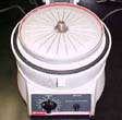
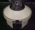
The packed cell volume (PCV) of blood is obtained by centrifuging a tube of blood and measuring the percentage of packed red blood cells (RBC’s) in the total sample. The blood is centrifuged (spun in an apparatus) and the PCV is read directly from a scale etched on the tube. If a microhematocrit centrifuge is used, heparinized capillary tubes (very small diameter tubes ~ 1 mm) are filled with blood and the ends are sealed. The capillary tubes are spun at high speed for 5 minutes and the PCV is read from a graph or chart. Determining the PCV is one of the best methods of detecting anemia (reduction in red blood cells).
RBC (Red Blood Cell Count)
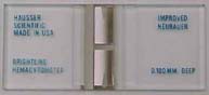
The RBC count is made after drawing either free-flowing blood or well-mixed blood containing an anticoagulant into an RBC pipette (an automatic machine such as a Coulter counter may also be used). The blood is then diluted with 0.85% NaCl (sodium chloride-salt) or another similar diluent. The blood is mixed by a mechanical shaker or by hand for 2 minutes. The fluid is used to fill a hemacytometer (thick glass slide specifically used for cell counts with a grid etched into the glass) and examined under a microscope. The rbc’s are then counted. A decrease in red cells is referred to as anemia. It may be due to decreased production of red cells by the bone marrow (hypoplastic or aplastic anemia) or to increased destruction of mature cells (hemolytic anemia). An increase in red cells is called polycythemia. It may be due to hemoconcentration or excessive production of RBC’s.
Blood Smear Examination of RBC’s
A blood smear is made by placing a small drop of fresh blood on a slide and backing another slide up to the drop and then quickly pulling the drop across the slide producing a thin film of blood with a feathered edge. A smear may be made with blood treated with heparin or EDTA soon after collection but it is preferable to make one with fresh blood. The slide is air-dried and then fixed in absolute methanol for one minute prior to staining. The staining procedure requires a quick dip in a fixative then dips in two separate staining jars, followed by washing. This procedure takes less than a minute. There are other methods of staining but this one is the most reliable and gives the best results.
- The smear is then examined under a microscope to evaluate the uniformity of the cells’ distribution and for possible detection of microfilaria (heartworm larvae). Clumps of tumor cells may also be detected at this time.
- Erythrocytes (RBC’s) are inspected for irregularities in size (anisocytosis). Smaller erythrocytes (microcytosis) and larger erythrocytes (macrocytosis) are noted. Variations in shape of the rbc’s is called poikilocytosis and should also be noted.
- Nucleated erythrocytes should be counted (indicative of anemia), Howell-Jolly bodies (thick, small, round remnants of nuclear DNA) may occasionally be seen in the cytoplasm of rbc’s and Heinz bodies (erythrocyte refractile bodies-cytoplasmic clumps of denatured hemoglobin) which are fairly common in cats should be noted. Heinz bodies are seen in cats that have been exposed to oxidants such as aspirin, and sulfonamides as well as certain plants. Cats should never have onions or garlic (which is in the onion family) since both cause Heinze body anemia.
- When reticulocytes (immature rbc’s that have lost their nuclei) are present in increased numbers, they reflect accelerated erythropoiesis (formation of red blood cells) and are associated with anemia.
- The smear should also be examined for evidence of blood parasites such as heartworms, and Haemobartonella.
- Many different anemias may be determined by examination of the rbc’s in the blood smear.
WBC (White Blood Cell Count)
The WBC count is made using free-flowing blood or well-mixed blood containing an anticoagulant and a WBC pipette. The blood is diluted in 0.1N HCl (hydrochloric acid), mixed well, and then used to fill a hemacytometer. A decrease in white cells may indicate the presence of a virus which can be as simple as a cold or as fatal as FIP (Feline Infectious Peritonitis). An increase in white cells usually indicates a bacterial infection. The severity of the increase or decrease of white cells can help to determine the severity of the condition and possible treatment.
Differential White Cell Count
A blood smear slide is prepared and a count is taken of the different type of white blood cells.
- In most species, the neutrophil is the most common white blood cell as it is in cats. Only mature neutrophils are present in the peripheral blood of normal animals. With staining, the granules in the cytoplasm stain pink and the nucleus stains a dark blue. In animals with bacterial infections, immature stages of the neutrophil are present in peripheral blood. They are called band cells.
- The eosinophil is similar to the neutrophil except that in staining, the cytoplasm contains red granules. Eosinophilia is indicative of parasitism or allergies.
- The basophil leukocyte contains large, coarse granules in its cytoplasm. These cells are seldom found in normal cats. Basophilia is seen in cats with toxemia (bacterial poisoning) or septicemia (disease causing bacteria in the blood).
- The lymphocyte is the next most common white blood cell found in most animals. It has a large, round, dark-staining nucleus and a small amount of pale blue cytoplasm. Lymphoblasts (cells which give rise to lymphocytes) detected in the blood indicate malignant disease of the lymphopoietic (lymphocyte forming) system. This includes Hodgkin’s disease, lymphosarcoma, lymphocytic leukemia, and lymphoma.
- The monocyte is larger than lymphocytes and mature neutrophils. It has a large quantity of pale blue cytoplasm that often contains vacuoles (clear space filled with fluid or air). An increase of monocytes could imply a viral infection is present.
Neutrophilia (increase in neutrophils=increase in wbc’s) may indicate bacterial infection, inflammation, toxemia, stress, exercise, or fear. Neutropenia (decrease in neutrophils=decrease in wbc’s) indicates shock. Neutropenia also occurs in cats during anesthesia. The cat’s body is able to respond rapidly to stress or disease by producing an increase or decrease in wbc’s within 3 to 4 hours of being affected and is the body’s natural defense mechanism to infection.
Blood Clotting Time
Coagulation (clumping of blood cells) disorders can be detected by measuring the clotting time or bleeding time. These tests are sometimes done before surgeries that may involve excessive loss of blood. Bleeding time can be determined by applying a thin layer of Vaseline to an area on the cat’s earflap, nose, lip, or footpad and cutting the skin deeply enough so that blood flows from the wound. Drops of blood are absorbed on a filter paper at 30 second intervals until the bleeding has stopped. The bleeding time should be between 1 to 3 minutes.
Clotting time can be measured in several different ways. One of the easiest is the capillary tube method. Freely flowing blood from a venipuncture (puncturing a vein such as with a needle) or nick in the skin is collected into capillary tubes (very small diameter tubes ~ 1 mm) without any anticoagulant. At 30 second intervals a segment of a capillary tube is broken off while checking closely for formation of a fibrin strand (filament that entangles rbc’s, wbc’s, and platelets to form a clot) between the pieces of the tube. This is continued until a fibrin strand is formed. The time that elapses from filling the tube with blood to formation of a fibrin strand is the clotting time. It normally takes 8 minutes in a cat.
Measurement of prothrombin (chemical substance that produces thrombin, which in turn produces fibrin) time provides information about the level of clotting factors. To measure prothrombin time, 9 parts of freshly drawn blood are mixed with 1 part of 3.8% sodium citrate solution. The mixture is centrifuged and the plasma is removed from the top. A 0.1 ml of the sample is transferred to a test tube and put in a 37°C water bath for one minute. Then 0.2 ml of commercial thromboplastin (clotting catalyst – factor III) is blown through a pipette into the plasma. The time required for visible clot formation is the prothrombin time. It normally takes 10 to 14 seconds. If the time is prolonged, there is either a deficiency in plasma prothrombin, fibrinogen (protein that is converted to fibrin through the action of thrombin when in the presence of calcium ions), or one of the clotting factors (V, VII, or X). A prothrombin time over 20 seconds usually is due to liver disease, warfarin (rat) poisoning, vitamin K deficiency, or a clotting factor deficit.
ESR (Erythrocyte Sedimentation Rate)
In general an accelerated ESR indicates disease. Some of the most common causes are inflammatory lesions with associated tissue damage, pregnancy, anemia, and neoplasia (development of a tumor or growth). Factors such as an increase in fibrinogen or serum globulins, a decrease in serum albumin, may cause an increase in the ESR. An increase in RBC counts and certain drugs may decrease the ESR. This test has most application to the dog, in which the normal ESR ranges from 4 to 10 mm/hour. The ESR is quite variable in the cat and of little value.
Platelet (Thrombocyte) Count
Using freshly drawn blood, mixed with a small amount of an anticoagulant such as EDTA, 13 µl of blood is mixed with 1.3 ml of 1% ammonium oxalate in a diluting pipette. The mixture is mixed thoroughly and allowed to stand for 10 minutes so that the rbc’s are completely lysed (destroyed). A hemacytometer is filled and the platelets which are smaller than rbc’s are counted.
When a small blood vessel is injured, platelets adhere to each other and to the edges of the injury and form a plug which covers the area. The plug or blood clot formed soon retracts and stops the loss of blood.
Thrombocytopenia (reduced platelet count) occurs in acute infections, anaphylactic shock (severe allergic reaction), certain hemorrhagic (bleeding) diseases, and anemias. Thrombocytosis (increased platelet count) occurs after operations, and following violent exercise and tissue injury.

Hemoglobin
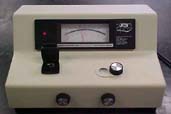
Hemoglobin is the iron-containing pigment of the red blood cells. Its function is to carry oxygen from the lungs to the tissues. Common methods of measuring hemoglobin include the use of a hemoglobinometer for oxyhemoglobin (the combined form of oxygen and hemoglobin which is found in arterial blood) and a spectrophotometer for cyanmethemoglobin (hemoglobin where the ferrous iron has been oxidized to ferric iron producing cyanosis, bluish or grayish discoloration of the skin). Using the hemoglobinometer, the chamber is filled with blood that is then mixed with a saponin-coated stick (unabsorbable glucoside (sugar) contained in roots of some plants) to lyse all of the rbc’s. When the chamber is then examined with transmitted light, two green fields are seen. The movable one is adjusted so that the colors of both fields match as closely as possible. The results obtained are reported as grams of hemoglobin per 100 ml of blood. The measurement of cyanmethemoglobin will give a slighly higher but more accurate result. The spectrophotometer is standardized with a hemoglobin standard and a blood sample is analyzed.
Mutated hemoglobin can cause severe hemolytic anemia which is usually fatal, hematuria (blood in the urine), including pain in bones, joints, abdomen, and chest. Cyanmethemoglobin causes cyanosis which is benign because of the presence of normal hemoglobin.
MCV (Mean Corpuscular Volume)
The MCV denotes the average size of erythrocytes. The MCV is determined by multiplying the PCV by 10 and dividing the product by the erythrocyte count per cubic millimeter.
MCH (Mean Corpuscular Hemoglobin)
The MCH denotes the average hemoglobin weight per red blood cell and is calculated by multiplying the hemoglobin in g/100 ml of blood by 10 and dividing the product by the erythrocyte count in millions per cubic millimeter.
MCHC (Mean Corpuscular Hemoglobin Concentration)
The MCHC is the average concentration of hemoglobin in red blood cells. The MCHC is determined by dividing the hemoglobin in g/100 ml with the PCV and multiplying by 100. The result is a percentage. The MCHC is a good indicator of hypochromic anemia (blood cells that have a reduced amount of hemoglobin). Hypochromic anemia may result from chronic bleeding or iron deficiency.
Normal Ranges and Means for Blood Values in Cats 1
| Component | Unit | Range | Mean (Avg) |
| RBC | cubic mm | 5.5 million-10 million | 7.5 million |
| Hemoglobin | g/dL | 8-15 | 12 |
| PCV | % | 24-45 | 35 |
| MCV | fL | 40-55 | 48 |
| MCHC | % | 30-35 | 33 |
| WBC | cubic mm | 8,000-25,000 | 17,000 |
| Differential Count | Unit | Range | Mean (Avg) |
| Neutrophil | % | 35-75 | 55 |
| Bands | % | 0-3 | 1.5 |
| Lymphocytes | % | 20-55 | 37.5 |
| Monocytes | % | 1-4 | 2.5 |
| Eosinophils | % | 2-12 | 7 |
| Basophils | % | rare | 0 |
Information on this site is for general information purposes only and is provided without warranty or guarantee of any kind. This site is not intended to replace professional advice from your own veterinarian and nothing on this site is intended as a medical diagnosis or treatment. Any questions about your animal’s health or diet should be directed to your veterinarian.
© 2022 Feline Nutrition Center. All Rights Reserved.
Privacy | Terms
